Isolation of cellular slime moulds
If you spread a very small amount of soil sample over an agar surface along with food bacteria and incubate the plate for 3 to 4 days, you will see growing hyphae of filamentous fungi and quite often fruiting bodies of cellular slime moulds as well. It is easier to isolate slime moulds on non-nutrient agar on which filamentous fungi do not grow fast.
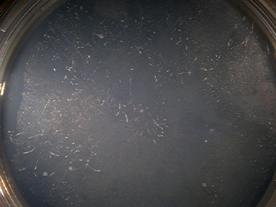
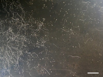
Three days after plating of a soil sample and bacteria. |
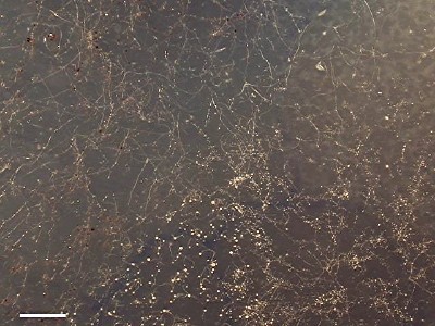
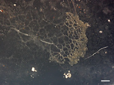
Seven days after plating (another example). |
A colony of P. pallidum. Slugs developing from aggregation streams can be seen below the centre and upper right corner of the photograph. The brownish white spots on the upper left and bottom left are colonies of a filamentous fungus. Five days after plating. Scale bar = 2 mm. (Kyoto, Japan). |
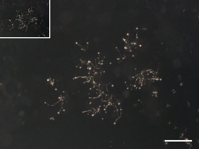
Small cellular slime mould (D. minutum?). Such small fruiting bodies could be easily overlooked. The inset shows the same one shown at the same magnification as the left photograph. Five days after plating. Scale bar = 1 mm. (Kyoto, Japan). |
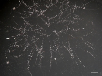
In the left is a colony of P. pallidum. To the right, many long and slender migrating slugs of D. polycephalum can be seen. Six days after plating. Scale bar = 5 mm. (Kanagawa, Japan). |
It is not common to see plasmodial slime moulds with such isolation methods. This picture shows a rare encounter of Physarum polycephalum and Dictyostelium polycephalum. Seven days after plating. Scale bar = 1 mm. (Kanagawa, Japan). |

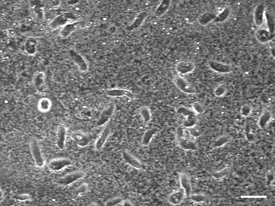
Nematodes often flourish on isolation plates. Scale bar (inset) = 0.1 mm. (Kyoto, Japan). |
On agar plates, ciliates look like large amoebae that do not change shape. Scale bar = 50 μm. (Kyoto, Japan). |